December 2024

Urinary system globeayush.com
Blog

Over view of the Urinary System: Embryology , Functions and Congenital Diseases

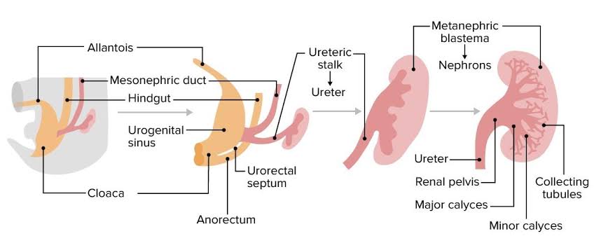
Introduction As we all know that nutrition is very essential to human body for its maintenance. Same as that body has to wash out all the waste and toxic substances which are produced after all the metabolic process , this work is done by excretory system also called as urinary system. COMPONENTS OF URINARY SYSTEM Development and Embryology of Urinary System This urinary system starts developing at 3rd week of Intrauterine life. This urinary system develops from the mesodermal ridge[Intermediate mesoderm] Bilaminar Disc Formation: The bilaminar disc forms during the 2nd week of embryonic development, following the process of implantation of the blastocyst into the uterine wall. Trilaminar Disc Formation: The trilaminar disc forms by the end of the 3rd week of development, during the process of gastrulation. Gastrulation is a crucial event that reorganizes the bilaminar disc into a three-layered structure, which will eventually give rise to all the organs and tissues of the body. Three divisions of Mesoderm: The Urinary System is mainly formed from the Intermediate Mesoderm. Formation of urinary system starts at week – 4 In 4th week of intra uterine life the intermediate mesoderm starts condensation and formation of nephrogenic cord and urogenital ridge takes place . Three overlapping kidney systems are formed from cranial to caudal region during the intra uterine life, they are : PRONEPHROS : These structures formed at the beginning of 4th week and these are rudimentary and non-functional occur in cervical region. These structures represented by 7 to 10 solid cell groups. These structures form vestigial excretory units called nephrotomes By the end of 4th week all indications of pronephric system have disappeared . MESONEPHROS : These structures are functional but for a short time , occurs in thoraco-lumbar region [upper L3 segments]. These mesonephros form mesonephric duct also called as wolffian duct and structures arise from this duct are called as mesonephric tubules. These mesonephros acts as primitive urinary system. Mesonephric duct which formed gets extended into cloaca and the mesonephric tubules form connections with the aorta through angiogenesis.[The process of formation of new blood vessels from existing blood vessel in body] NOTE : From the aorta the waste gets collected into the mesonephric tubules and from there through mesonephric duct they passed into cloaca .Cloaca is the structure which collects both urine and feces and send them to allantois , this is the structure which get modified into the anus and urinary bladder and urethra in later stage. These mesonephros works as primitive urinary system from week 5 to week 10 of intra uterine life . METANEPHROS : These structures are functional and permanent and forms kidney , these appear at 5th week. These metanephros are formed in pelvic region. The metanephros starts secreting growth factors which acts on mesonephric duct [wolffian duct] and induce formation of ureteric bud. Several important growth factors are involved in the interaction between the metanephros and ureteric bud: RECIPROCAL INDUCTION The ureteric bud (UB) plays a critical role in kidney development through a process called reciprocal induction, where it interacts with the surrounding metanephric mesenchyme to drive the formation of the functional structures of the kidney. Reciprocal induction refers to the mutual signaling between two different tissues to promote the development of one another. In the case of kidney development, the ureteric bud and metanephric mesenchyme interact in such a way that each tissue influences the development of the other. Reciprocal induction that works between the ureteric bud and the metanephric mesenchyme: 1. Ureteric Bud Signals to the Metanephric Mesenchyme The ureteric bud, once it outgrows from the mesonephric duct (Wolffian duct), interacts with the metanephric mesenchyme and induces it to undergo a process called mesenchymal-to-epithelial transition (MET). This is essential for the formation of nephrons . 2. Metanephric Mesenchyme Signals to the Ureteric Bud In response to signals from the ureteric bud, the metanephric mesenchyme also sends signals back to the ureteric bud to control its growth and branching pattern. This reciprocal signaling is essential for the proper formation of both the nephron and the collecting system. Formation Of Renal Pelvis And Other Structures The uretic bud which extends into the metanephric blastomere through ureteric stalk forms renal pelvis . Renal pelvis form extensions and formation of major calyces takes place , these major calyces gives rise to minor calyces .At the ends of minor calyces origin of collecting tubules takes place[ approximately 1 to 3 million tubules]. The collecting tubules which are formed release growth factors and helps in condensation of metanephric blastomere at the place where it surrounds the collecting tubule to form metanephric mesodermal cap. This metanephric mesodermal cap get modified into metanephric vesicle and then into metanephric tubule and get fused with the collecting tubule to form distal convoluted tubule [DCT] , it goes on modifying and form proximal convoluted tubule [PCT] and bowman’s capsule respectively. The abdominal aorta divides into two branches at the pelvic region into right and left iliac arteries these form tuft of blood capillaries which form glomerular capillaries into bowman’s capsule of the nephron. During this stage the metanephric mesoderm deepens between the PCT and DCT which form loop of Henle. Hence , kidney develops from 2 sources : Position of kidney Initially it is situated in pelvic region later it shifts to more cranial position in the abdomen . In pelvic region the metanephros receive blood supply from the pelvic branch of aorta which gradually gets degenerated . During ascent to abdominal level, it is vascularized by arteries that originated from the aorta at continuously higher levels .The lower vessels gradually get degenerated ,at last kidneys are supplied by renal arteries which are developed lateral branches of aorta. Functions of Kidney The metanephros completely becomes functional at week 12 and take over all functions of kidney and mesonephric duct no longer plays any role in formation of urine it takes crucial role in formation of genital tract. Urine passed into the amniotic cavity and get mixes with the amniotic fluid

Urine Examination: Modern and Ayurvedic Aspects
Blog

Urine Examination: Modern and Ayurvedic Aspects

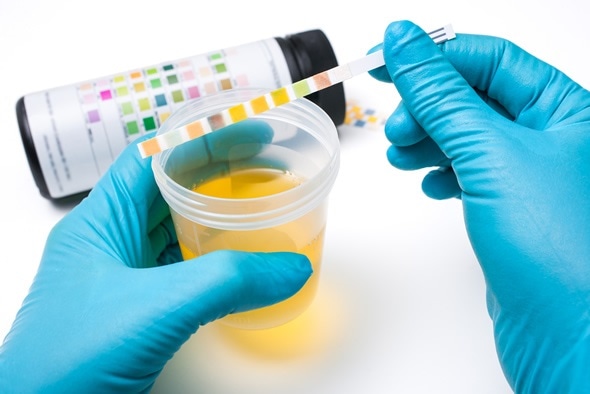
Introduction Urine examination, known as urinalysis, is one of the most commonly used diagnostic tools in modern medicine. It evaluates physical, chemical, and microscopic properties of urine to assess overall health, detect diseases, and monitor treatment efficacy. Ayurveda, the ancient Indian medical science, also places significant importance on urine examination, referring to it as Mutra Pareeksha for diagnosing imbalances in doshas (Vata, Pitta, and Kapha) and metabolic disorders. In this article compre we will explores urine examination from both modern and Ayurvedic perspectives, integrating their diagnostic methodologies and clinical applications. Anatomy and Physiology of the Urinary System The urinary system comprises the kidneys, ureters, urinary bladder, and urethra, all of which work together to filter blood, eliminate waste, and maintain homeostasis. In Ayurveda, the urinary system is governed by Mutravaha Srotas and is closely associated with Apana Vata, the sub-dosha responsible for excretion. Modern Aspects of Urine Examination Types of Urine Tests 1. Physical Examination: 2. Chemical Examination: Utilizes dipstick tests for detecting: 3. Microscopic Examination: Applications in Modern Medicine Ayurvedic Aspects of Urine Examination Historical Context Ayurveda emphasizes urine examination through Mutra Pareeksha, as elaborated in classical texts like Charaka Samhita, Sushruta Samhita, and Ashtanga Hridaya. It identifies metabolic imbalances and provides clues about systemic diseases. Methodology of Mutra Pareeksha Parameters Evaluated in Ayurveda Applications in Ayurvedic Medicine Analysis: Modern vs Ayurvedic Perspectives Aspect Modern Medicine Ayurveda Focus Organ-specific and systemic health Dosha imbalance and metabolic health Techniques Physical, chemical, and microscopic Observation and dosha-based analysis Disease Detection Infections, diabetes, kidney issues Prameha, Mutrakricchra, Ashmari Technology Advanced lab methods and imaging Tailabindu Pareeksha, sensory analysis Approach Objective and measurable Holistic and individualized Case Studies Case 1: Diabetic Patient Case 2: Urinary Tract Infection Limitations and Integration Modern Limitations Ayurvedic Limitations Integration Opportunities The integration of modern and Ayurvedic urine examination offers opportunities to enhance diagnostic accuracy, enable early detection, and provide personalized care. Modern tools ensure precision, while Ayurveda focuses on holistic evaluation and root-cause analysis through dosha-based methods like Tailabindu Pareeksha. Combining both systems allows for comprehensive disease management, especially for chronic conditions like diabetes and kidney disease, by blending advanced treatments with Ayurvedic lifestyle, dietary, and herbal interventions. This approach is cost-effective, promotes research on Ayurvedic methods, and fosters innovation in healthcare, bridging ancient wisdom with modern science. Conclusion Urine examination remains a cornerstone in both modern and Ayurvedic diagnostics. While modern medicine offers detailed biochemical and microscopic analysis, Ayurveda provides a holistic framework based on dosha evaluation. Together, they represent a complementary approach to understanding health and diagnosing diseases. By integrating the strengths of both systems, practitioners can offer more comprehensive care, emphasizing early detection, prevention, and individualized treatment. References Read other articles

Aragwada Ayurvedic drug : Classical and Modern Aspects , Research
Blog

Aragwada Ayurvedic drug : Classical and Modern Aspects , Research

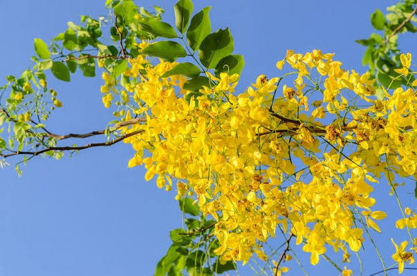
Basonym of Drug आ समन्ताद् रुजां वधोऽत्र । Aragwadha completely cures ailments. Botanical Name: Cassia fistula Linn. Cassia-Kasia (Greek), derived from a Hebrew word, which  is  a name of plant.fistula-Tube (Refering to the cylindrical pod)Family-Fabaceae (See appendix-II for family features). Classification of the Dravya Charaka Kushthaghna, Kandughna Varga Sushruta Aragwadhadi gana, Shyamadi ganaB.P.Ni.Haritakyadi varga. When flowering, the tree looks adorned with garlands. Aragwadha fruit has depression at every 4 inch distance. Aragwadha fruits are long. Aragwadha tree is regarded as king of tree oving to its beauty. Aragwadha flowers are golden yellow coloured. Aragwadha tree is liked by people as it is beautiful. Properties and actions Aragwadha relieves Ama Expels mala (faeces) from the body. Pods are therapeutically very useful. Aragwadha cures skin diseases. Aragwadha relieves from fever. Aragwadha is useful in many diseases. Others Aragwadha corrects the digestive process. Aragwadha flowers were used as ear ornaments. RASA PANCHAKA : Rasapanchaka Phalamajja Rasa Madhura, Tikta Guna guru, snigdha Virya sita Vipaka madura DOSHA KARMA. Karma (Actions)Phala (Fruit) – Sramsana, Shulahara, Ruchya, Koshtha Shuddhikara, Krimighna, Pramehaghna, Jwaraghna, Kushtaghna Patra (Leaf)-Meda vishoshaka, Virechaka.Pushpa (Flower) – GrahiAragwadha phala majja is best Sramsaka. Dosha karma :Vatapitta Shamaka and Pitta kapha samshodhaka.Vata Shamaka because of Madhura rasa, Snigdha guna and Madhura vipaka.Pitta Shamaka because of Madhura rasa and Shita virya.Pitta kapha, Samshodhaka because of its Sramsana property. Agrya Karma चतुरङ्गलो मृदु विरेचनानाम् । (च.सू. 25/40)Chaturangula (Cassia fistula Linn.) induce mild purgation Pharmacological actions • Purgative• Feverish Rogaghnata (Therapeutic indications) Phala (Fruit)-Jwara, Hradroga, Raktapitta, Udavarta, Kushtha Krimi Prameha, Mutrakrachra, Gulma, Udara, Vrana. Patra (Leaf) Medoroga Amayika prayoga (Therapeutic administration) हरिद्रा मेहिनं राजवृक्षकषायम् । (सु.चि. 11/9) Aragwadha (Cassia fistula) kashaya is best for Haridra meha. पर्णानि पिष्ट्वा चतुरङ्गुलस्य तक्रेण पर्णान्यथ काकमाच्याः । तैलक्तगात्रस्य नरस्य कुष्ठान्युद्वर्तयेद्‌वहनच्छदैश्च ।। (च.सू. 3/17) Triturate leaves of Aragwadha (Cassia fistula) and Kakamachi (Solanum nigrum) with buttermilk. The paste thus obtained is applied over skin lesions (the part should be anointed prior to this application). आरग्वधस्य पत्राणि भृष्टानिकटुतैलतः । आमघ्नानि नरः कुर्यात् सायं भक्तावृताति च ॥ (भा.प्र.चि. 26/52) Leaves of Aragwadha (Cassia fistula) are fried with mustard oil. These leaves are chewed after evening meal. आरग्वधस्य पत्राणि त्वचं श्लेष्मातकस्य च। पृथगालेपनं कुर्यात् द्वन्द्वशः सर्वशोऽपि वा । प्रदेहाः सर्व रावैते देयाः स्वल्पघृताप्लुताः ।। (च.चि. 21/89-92) Paste of Aragwadha (Cassia fistula) leaves and Shleshmataka bark is mixed with ghee and applied in all types of Visarpa (Erysipelas). Matra (Dose) Bija churna (Powder);3_6 g Mula twak kwatha;10-15ml Pushpa swarasa 5-10 ml Phala majja (Fruit pulp) 10 to 20 g Important phytoconstituents Pod pulp – Anthraquinone, sucrose, lignoceric acid, amino acids. Root bark – Anthraquinone derivates, sennoside A&B, Fistucacidin, g-sitosterol, Volatile oil. Stem bark and Leaves – Rhein glycoside. Rogaghnata (Therapeutic indications) Phala (Fruit)-Jwara, Hradroga, Raktapitta, Udavarta, Kushtha Krimi Prameha, Mutrakrachra, Gulma, Udara, Vrana FORMULATIONS & Indication Aragwadhadyarista- jwara, hrdroga, vatarakta Aragwadhadi Avaleha-Udavartha, hrdroga Aragwadhadi ghrta- jwara, vatarakta Aragwadhadi kashaya-sula, kamala, raktapitta Aragwadhadi taila- switra Mahamarichyadi ttaila-kustha, sarvanga, sotha. Habitat Grows all over India and as it is a beautiful plant, it is cultivated in gardens. External Morphology Habit-A medium sized perennial tree.Stem-Erect, branched, cylindrical, woody, solid, bark greenish grey in colour.Leaf-Alternate, petiolate, compound, paripinnate, with pulvinus base, leaf lets 4-8 pairs, stalked, ovate, wavy, acute, glabrous, entire, venation of the leaflets unicostate, reticulate.Inflorescence-Long axillary pendulous racemes.Flower Pedicellate, with long pedicels, bracte- ate, bisexual, complete, zygomorphic, yellow, hypogy- nous and pentamerous.Calyx-5 speals, slightly petaloid, polysepalous, odd sepal anterior, imbricate aestivationCorolla-5 petals, polypetalous, yellow, posteriorpetal is the small and innermost in the bud; petals unequally inserted on the thalamus.Androecium-10 stamens, polyandrous and all with anthers, but very unequal in length.Gynoecium-Monocarpellary, ovary superior, unilocular, with marginal placentation, ovules many, short style, stigma is terminal and hairy.fFruit-A legume which is very long (1-2 feet) and cylindrical (nearly one inch in diameter).Seeds-Flat, albuminous, embedded in soft pulp.Varieites :Dhanvantari Nighantu & Raja Nighantu: 1. Aragwadha 2. Karnikara Research Corner: Gallery FRUIT OF ARAGWADA. FLOWER OF ARAGWADA. LEAF OF ARAGWADA Read other articles

Kajjali globeayush.com
Blog

Kajjali in Ayurveda: Preparation, Benefits, Key Formulations, and Applications

Introduction Kajjali, also known as black mercury sulfide, is a crucial preparation in Rasashastra, a specialized branch of Ayurveda. Rasashastra primarily involves therapeutic uses of mercury (Parada) and other minerals. While mercury in its natural form is toxic and unsuitable for consumption, Ayurveda processes it into a safe and effective compound, Kajjali, by triturating it with sulfur (Gandhaka). This article explores Kajjali’s preparation, properties, and its versatile applications in Ayurvedic medicine. Definition and Significance of Kajjali Kajjali is prepared by grinding purified mercury with sulfur until the mixture achieves a fine black, smooth texture devoid of shiny particles. It is extensively used in Rasashastra because it: Historical Background of Kajjali Kajjali finds its roots in ancient Ayurveda, where the transformation of raw mercury into medicinal forms was first documented by Acharya Nagarjuna in the 8th century. Its therapeutic uses expanded with time, gaining recognition in formulations like Rasa Parpati and Arogyavardhini Vati. Despite its therapeutic potential, it became popular in Europe only by the 10th century but remained less understood for its medicinal applications. Why is Kajjali Significant in Ayurveda? Preparation of Kajjali Purification Process To eliminate toxicity, mercury and sulfur undergo a rigorous purification process: Trituration Process (Mardana Sanskara) The purified mercury and sulfur are triturated in a mortar and pestle: Ratios of Mercury and Sulfur in Kajjali Different therapeutic applications require varying proportions of mercury and sulfur: Before use, Kajjali is subjected to rigorous testing: Utility of Kajjali in Rasashastra Kajjali is used as: Applications of Kajjali Therapeutic Applications of Kajjali General Properties Ratio Therapeutic Uses Sahapana (Adjunct) 1:1 Vomiting, boils, indigestion Honey, herbal decoctions 1:2 Chronic skin conditions Ghee, warm water 1:3 Respiratory disorders Milk, Tulsi juice Ratios and Their Applications Kajjali’s applications depend on the ratio of mercury to sulfur. Ratio Examples of Rasayogas Therapeutic Uses 1:1 Mrutyunjaya Rasa, Arogyavardhini Vati Treating fever, digestive issues 1:2 Rasaparpati, Jeevananama Rasa Respiratory disorders, skin issues 1:3 Triguna Gandhaka Kajjali, Rasasindhura Chronic diseases, vitality booster 1:4 Mahaveerari Rasa, Udaya Bhaskara Rasa Severe skin conditions, immunity Formulations with Indications Formulation Type of Kajjali Indication Adjunct (Anupana) Arogyavardhini Vati Samaguna Kajjali Liver disorders, skin diseases Warm water Rasasindhura Triguna Kajjali Chronic conditions, rejuvenation Honey and ghee Priyangu Kajjali Samaguna Kajjali Jwara (fever), Raktapitta Honey Kutaj Kajjali Samaguna Kajjali Chronic diarrhea, dysentery Warm water Pushkarmoola Kajjali Samaguna Kajjali Respiratory distress Tulsi decoction Madhuyashti Kajjali Samaguna Kajjali Cough, headache, sinusitis Licorice decoction Detailed Indications Disease Recommended Kajjali Dose Adjunct Indigestion Samaguna Kajjali 120 mg Lemon juice, dry ginger powder Skin disorders Dwiguna Kajjali 240 mg Butter or neem oil for topical application Asthma and bronchitis Dwiguna Kajjali 480 mg Tulsi juice, honey Alcoholism Samaguna Kajjali 240 mg Amla juice and sugar Cervical lymphadenopathy Samaguna Kajjali Local application Calotropis latex Syphilis Dwiguna Kajjali 360 mg Cow butter List of Kajjali-Based Formulations Name Disease Indication Type of Kajjali Kajjali Content (per dose) Arogyavardhini Vati Skin disorders, liver diseases Samaguna Kajjali 5.5% Icchabhedi Ras Digestive issues Samaguna Kajjali 25% Chandrakala Ras Urinary disorders Dwiguna Kajjali 20% Trimurti Ras Obesity, metabolic disorders Samaguna Kajjali 66.66% Hridayarnav Ras Cardiac disorders Samaguna Kajjali 66.66% Lakshmi Vilas Ras Fevers Samaguna Kajjali 20% Modern Scientific Insights Chemical Composition Pharmacological Properties Toxicity Studies Unique Features Advanced Properties and Modern Analysis Conclusion Kajjali exemplifies the sophistication of Ayurvedic alchemy, transforming toxic mercury into a safe, therapeutic agent. Its versatile applications, from treating chronic diseases to enhancing immunity, demonstrate its indispensability in Ayurveda. As modern research continues to validate its efficacy and safety, Kajjali remains a cornerstone in bridging ancient wisdom with contemporary healthcare. References Read other articles

Comprehensive View Of Pericardium: Understanding Anatomy, Function, Development and Congenital Anomalies ,Clinical Significance
Blog

Pericardium: Understanding Anatomy, Function, Development and Congenital Anomalies ,Clinical Significance

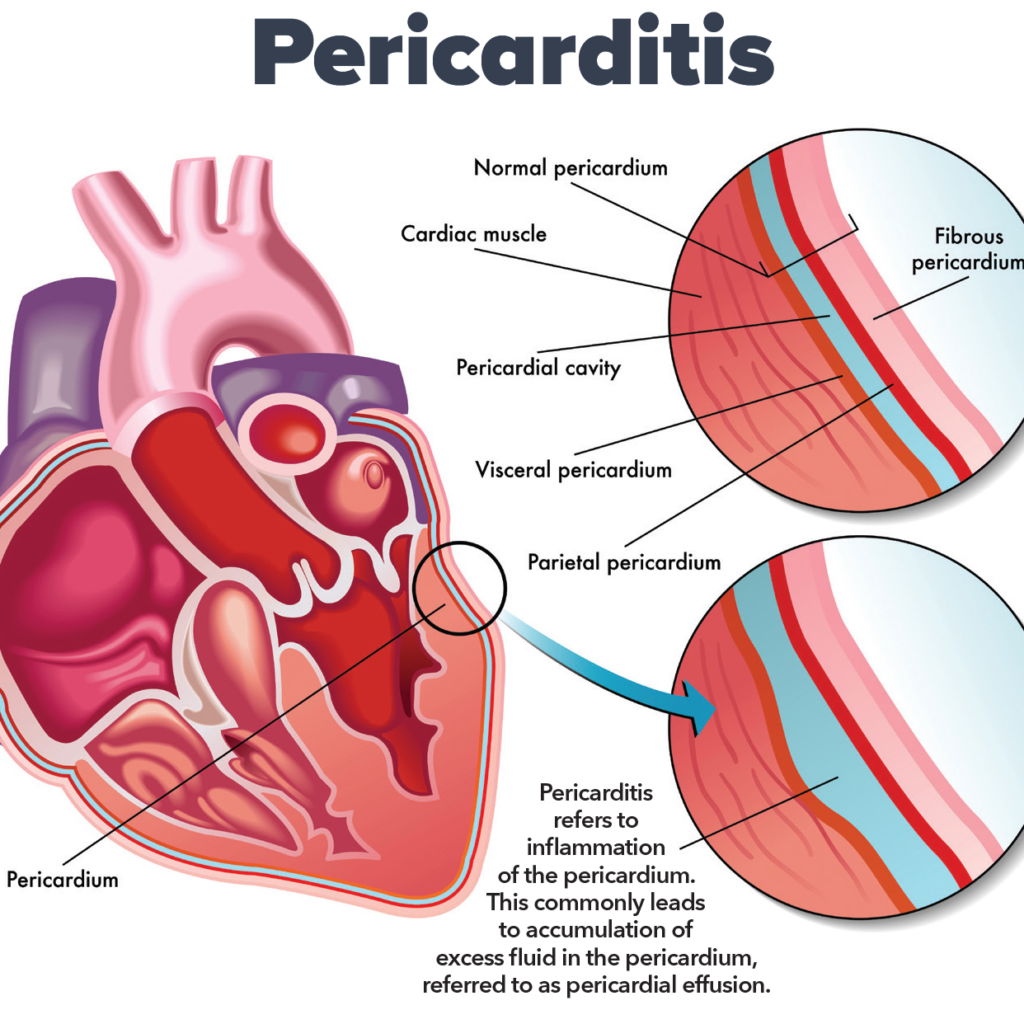
INTRODUTION: What is pericardium ? Term pericardium is derived from the Greek prefix :peri means – Around and Kardia means- Heart pericardium is a double walled fibro serous sac which encloses the heart and roots of great blood vessels. It is situated in middle mediastinum. It consists of 2 layers , they are as follows : STRUCTURE AND FUNCTIONS OF PERICARDIUM: 1.Fibrous Pericardium It is the outer most layer , which encloses the heart and fuses with the roots of great vessels which enter or leave the heart .It is conical sac and made up of dense and loose connective tissue, mainly collagen and elastin .It is non pliable [inflexible] , it can change some what. 1.1 Relations Of Fibrous Pericardium APEX : It is blunt, lies at the level of sternal angle [ angle of louis ] . It fuses with the roots of great vessels and with the pre tracheal fascia. BASE: Broad and inseparably blended with central tendon of the diaphragm. ANTERIOR: It is connected to upper and lower ends of body of sternum [breast bone] by weak superior and inferior sternopericardial ligaments. POSTERIOR It relates to: Each side 1.2 Functions : Protection: As it is made up of dense fibrous tissue it cushions the heart from outside forces and sudden pressure changes . Anchorage: As it is shows attachments with the diaphragm and sternum [ breast bone] it keep the heart in place. Restricting the heart volume : As it is made up of fibrous dense connective tissue .It prevents heart from more expansion than needed. Fibrous prolongations of pericardium: Vessels like aorta, superior vena cava, right and left pulmonary arteries and 4 pulmonary veins receives prolongations of fibrous pericardium up to 5 to 6 mm NOTE: Inferior vena cava doesn’t receive any covering from fibrous pericardium , it enters the pericardium through the diaphragm’s central tendon Protection from infections : Protects heart from infections that might spread from nearby organs like lungs. 2. Serous pericardium : It is a thin double layered serous membrane made up of mesothelium. Parietal layer : This layer fused with the inner surface of fibrous pericardium. Visceral layer: This layer fuses with the heart , except at the cardiac grooves. These two layers, parietal and visceral pericardium continues with each other at roots of great vessels. Pericardial cavity: The space between the parietal and visceral layers is called as pericardial cavity It is filled with pericardial fluid [serous fluid] –15 to 50 ml which is secreted by serous layer. 2.1 Functions: Secretion Of Pericardial Fluid: SINUSES OF PERICARDIUM: Sinuses are the extensions of the pericardial cavity that form between the pericardium and heart surface. There are 3 types of sinuses : TRANSVERSE SINUSES : It is located behind the aorta and pulmonary trunk, and in front of superior vena cava . horizontal gap between arterial and venous ends of heart tube Bounded by : OBLIQUE SINUSES :It is j-shaped narrow space located behind the heart , particularly the left atrium. Bounded by : IMPORTANCE : It permits pulsations of left atrium to take place freely. SUPERIOR SINUSES: It is located in front of ascending aorta and the pulmonary trunk NOTE: it can’t be accessed during electrophysiology procedures. CONTENTS OF PERICARDIUM : BLOOD SUPPLY : Fibrous and parietal layers are supplied by branches of: NERVE SUPPLY: DEVELOPMENT AND CONGENITAL ANOMALIES OF PERICARDIUM: Pericardium embryological development is a complex process that involves several stages, which are intricately linked to the formation of the heart and the cardiovascular system. The pericardium forms from mesodermal layers during embryonic development and undergoes key changes as the heart evolves into its mature anatomical state. 1. Embryological Origins of the Pericardium The pericardium develops from two main mesodermal sources: the somatic mesoderm and the splanchnic mesoderm. The space between these two layers, called the pericardial cavity, is initially a small slit, which later expands and houses the heart. 2. Stages of Pericardium Formation 2.1. Primitive Heart Tube Development (Week 3) At around the third week of embryonic development, the heart begins as a simple, tubular structure formed from the cardiogenic mesoderm. This tube undergoes folding and looping to form a more complex structure, eventually forming the heart with the atria and ventricles. Simultaneously, the pericardial cavity begins to form. Initially, the heart is surrounded by a small amount of mesodermal tissue derived from the lateral mesoderm. The pericardium begins as a continuous sheet of mesoderm surrounding the heart tube. As the heart tube folds, this mesoderm differentiates into the somatic mesoderm (which will form the parietal pericardium) and the splanchnic mesoderm (which will form the visceral pericardium). 2.2. Formation of the Pericardial Cavity (Week 4) By the fourth week, the pericardial cavity becomes more defined. The space between the parietal and visceral layers of mesoderm get enlarged and filled with fluid, allowing the heart tube to move freely within this sac. This fluid-filled pericardial cavity acts as a cushion, reducing friction as the heart begins to beat. 2.3. Development of Pericardial Layers (Week 5 to 6) During weeks 5 and 6, the pericardial cavity enlarges, and the pericardial layers become distinct. The visceral pericardium becomes intimately associated with the developing myocardium (heart muscle), and the parietal pericardium becomes a separate layer, lining the pericardial cavity. At this stage, mesodermal cells in the region of the epicardium (visceral pericardium) also begin to differentiate into epicardial cells, which will contribute to the development of the heart’s coronary vasculature. Meanwhile, the parietal pericardium develops a fibrous structure that will become the fibrous layer of the adult pericardium. 3. Maturation and Differentiation 3.1. Formation of the Fibrous Pericardium The fibrous pericardium is derived from the surrounding mesodermal tissues and becomes an important structure as the heart grows. During the later stages of embryonic development (by week 7-8), the parietal pericardium becomes organized into the fibrous pericardium, which is a tough, inelastic membrane that limits excessive movement of the heart and helps anchor it within the thoracic cavity. 3.2. Pericardial Fluid

Tridosha globeayush.com
Blog

Understanding Concepts 3 Doshas in Ayurveda: Tridosha

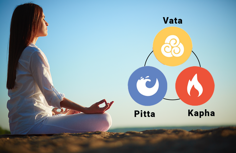
1. Introduction Significance of Tridosha in Ayurveda Tridosha, the three fundamental bio-energies—Vata, Pitta, and Kapha—forms the cornerstone of Ayurvedic medicine. It represents the biological energies derived from the five elements (Pancha Mahabhutas: Akasha, Vayu, Agni, Jala, and Prithvi). These energies govern all physiological, psychological, and pathological processes in the human body. Tridosha is the foundation for diagnosing, preventing, and managing diseases in Ayurveda, as it emphasizes restoring balance rather than merely treating symptoms. When in equilibrium, Vata ensures proper movement, Pitta maintains metabolism, and Kapha provides stability. Any disturbance in these doshas leads to disease, showcasing their vital role in maintaining health and wellness. Historical background and foundational principles The concept of Tridosha dates back to the Vedic era, with its earliest mention in texts like the Charaka Samhita and Sushruta Samhita. According to Ayurveda, life is a dynamic interplay of three forces—Vata (wind), Pitta (bile), and Kapha (phlegm)—that work in harmony to sustain life. Each dosha is associated with specific qualities (gunas) and functions. Ancient sages identified how these energies are influenced by diet, lifestyle, seasonal changes, and mental states, creating a unique, personalized approach to health care. The Tridosha theory is based on the idea that the body, mind, and environment are interconnected, and maintaining balance between the doshas leads to optimal well-being. Relevance of Tridosha in modern healthcare In modern healthcare, the Tridosha concept offers a holistic and preventive approach that aligns with personalized medicine. By recognizing individual constitution (Prakriti), Ayurveda provides tailored recommendations for diet, lifestyle, and treatment. Research has highlighted similarities between dosha imbalances and chronic diseases, such as metabolic disorders, stress-related illnesses, and inflammatory conditions. Additionally, the circadian rhythms of doshas correlate with modern chronobiology, emphasizing the role of time in physiological functions. With the growing emphasis on integrative medicine, Tridosha’s principles are gaining recognition for their focus on balance, natural therapies, and promoting long-term health through sustainable lifestyle practices. 2. Nirukti (Etymology) of Tridosha दूषयन्ति इति दोषाः । (A.H.Su. 1:6, Arunadatta) The literary meaning of the word dosha is that which causes decay, destruction or vitiates (others when in aggravated condition). However this is not the correct definition as we all know that when doshas are in balanced condition they carry out all prominent physiological function and maintain health. When doshas get disturbed, they attack other body tissues which are weak and disease is created. The elements, which get spoiled due to vitiated doshas are called as dushya (victims). They are dhatu (body tissue), mala (wastes), organs, channels, sense organs etc. In short dushya are everything in body excluding doshas. Knowledge about dosha is very fundamental in the process of learning Ayurveda. Concept of Tridosha has gradually evolved in Ayurveda, for example – 3. Definition of Tridosha Classical Ayurvedic Definitions The term “Tridosha” is derived from the Sanskrit words “Tri,” meaning three, and “Dosha,” meaning fault or defect. In Ayurvedic texts, “Dosha” is described as a dynamic principle responsible for both the maintenance and disruption of health. According to the Charaka Samhita, Tridosha governs all physiological and psychological functions. Each dosha is made of two primary elements: The equilibrium (samyavastha) of these doshas ensures health, while their imbalance (vikriti) causes disease. Functional Role of Doshas in the Human Body Each dosha plays a distinct role in the functioning of the body: 4. Tridosha: The Three Pillars of Life Tridosha forms the core of Ayurveda and symbolizes the three energies essential for life: Vata, Pitta, and Kapha. These energies control all functions of the body and mind, working together to maintain balance. Vata Dosha: The Energy of Movement Vata represents motion and is linked to air and space. It controls breathing, blood circulation, and all physical and mental movements. When balanced, it supports creativity and flexibility; when imbalanced, it causes restlessness and dryness. Pitta Dosha: The Energy of Transformation Pitta is the energy of heat and digestion, connected to fire and water. It governs metabolism, temperature, and mental focus. Balanced Pitta leads to sharp intellect and a healthy digestive system, but an imbalance can cause anger and inflammation. Kapha Dosha: The Energy of Structure Kapha provides stability and nourishment, linked to earth and water. It maintains body strength, immunity, and emotional calmness. In balance, it ensures growth and stability, while imbalance results in sluggishness and weight gain. 5. Subtypes of Doshas Subtype Specific Function Prana Vata Governs breathing, swallowing, thinking, and sensory perception. Udana Vata Controls speech, memory, energy, and upward movements like exhalation. Samana Vata Regulates digestion, assimilation, and movement of nutrients in the gastrointestinal tract. Vyana Vata Manages circulation, heartbeat, and overall body movements. Apana Vata Oversees elimination processes, such as urination, defecation, and childbirth. Subtypes of Pitta Dosha Subtype Specific Function Pachaka Pitta Responsible for digestion and metabolism in the stomach and intestines. Ranjaka Pitta Influences liver and spleen functions, maintaining blood quality and color. Sadhaka Pitta Supports intelligence, memory, emotional stability, and decision-making. Alochaka Pitta Controls vision and maintains eye health. Bhrajaka Pitta Regulates skin health, complexion, and temperature control. Subtypes of Kapha Dosha Subtype Specific Function Avalambaka Kapha Provides structural stability, supports heart and lung functions. Kledaka Kapha Protects the stomach lining, aids digestion, and maintains lubrication. Bodhaka Kapha Regulates taste perception and saliva production in the mouth. Tarpaka Kapha Nourishes and lubricates the brain and spinal cord for better mental health. Shleshaka Kapha Lubricates joints, supports smooth movement, and strengthens the skeletal system. 6. Tridosha in Diagnosis Diagnosis of Tridosha Dosha Signs of Imbalance Vata Dry skin, constipation, anxiety, irregular sleep, feeling cold. Pitta Irritable, inflammation, acidity, excessive heat, red complexion. Kapha Lethargy, excess weight, congestion, water retention, sluggish digestion. 7. Scientific Correlations with Tridosha Modern Interpretations of Dosha Functions While Ayurveda is an ancient system of medicine, its principles resonate with contemporary scientific understanding, particularly in the realm of body systems and functions. The doshas—Vata, Pitta, and Kapha—can be understood through modern biological systems and physiological processes. For example, Vata corresponds to the nervous system and movement in the body, as it governs all motion-related functions

Surya Namaskar globeayush.com
Blog

12 Asanas in Surya Namaskar: Modern aspect, anatomy

Surya Namaskar Asanas Surya Namaskar, or Sun Salutation, is a sequence of 12 asanas (poses) that integrate physical movement with controlled breathing. Below is a detailed description of each asana with its associated muscles, nerves, blood supply, and movements. 1. Pranamasana (Prayer Pose) Movement:Standing posture with palms together in Anjali Mudra. Primary Muscles:Rectus abdominisQuadriceps femorisErector spinae Nerves:Lumbar plexus, femoral nerve. Blood Supply:Descending aorta branches, femoral arteries. Involved Movements:Minimal movement; focus on balance and alignment.Contraction of abdominal muscles for upright posture. 2. Hasta Uttanasana (Raised Arms Pose) Movement:Arms stretched overhead, slight backward bend. Primary Muscles:Deltoids TrapeziusLatissimus dorsiErector spinae Nerves:Thoracodorsal nerve, spinal accessory nerve. Blood Supply:Subclavian and axillary arteries. Involved Movements:Extension of the thoracic spine.Flexion at the shoulder joint. 3. Uttanasana (Standing Forward Bend) Movement:Forward fold at the hips, touching the ground. Primary Muscles:Hamstrings (biceps femoris, semitendinosus, semimembranosus)GastrocnemiusErector spinae Nerves:Sciatic nerve, tibial nerve. Blood Supply:Popliteal artery, posterior tibial artery. Involved Movements:Flexion at the hip joint.Extension at the knee joint. 4. Ashwa Sanchalanasana (Equestrian Pose) Movement:One leg extended back, other knee bent. Primary Muscles:Hip flexors (iliopsoas)QuadricepsGluteus maximusSoleus Nerves:Femoral nerve, sciatic nerve. Blood Supply:Femoral artery, popliteal artery. Involved Movements:Extension of the hip and knee.Stabilization of the pelvic region. 5. Dandasana (Plank Pose) Movement:Body straight in a plank-like posture. Primary Muscles:Core muscles (rectus abdominis, transverse abdominis)Pectoralis majorDeltoids Nerves:Thoracoabdominal nerves, brachial plexus. Blood Supply:Axillary and subclavian arteries. Involved Movements:Isometric contraction of core.Stabilization of shoulder girdle. 6. Ashtanga Namaskara (Eight-Limbed Pose) Movement:Lowering the body, touching chest, knees, and chin to the floor. Primary Muscles:Pectoralis majorTriceps brachiiRectus abdominis Nerves:Median and ulnar nerves. Blood Supply:Subclavian and intercostal arteries. Involved Movements:Flexion at the elbow joint.Activation of abdominal muscles. 7. Bhujangasana (Cobra Pose) Movement:Upper body lifted, supported by palms. Primary Muscles:Erector spinaeSerratus anteriorDeltoids Nerves:Dorsal rami of spinal nerves. Blood Supply:Intercostal arteries, lumbar arteries. Involved Movements:Extension of the thoracic spine. Stabilization of shoulders. 8. Adho Mukha Svanasana (Downward-Facing Dog Pose) Movement: Body forms an inverted “V” shape. Primary Muscles:HamstringsTriceps surae (gastrocnemius and soleus)Erector spinae Nerves: Sciatic nerve, tibial nerve. Blood Supply: Popliteal artery, posterior tibial artery. Involved Movements:Extension at the hips. Stabilization of the shoulder girdle. 9. Ashwa Sanchalanasana (Equestrian Pose) (Repeat of step 4) 10. Uttanasana (Standing Forward Bend) (Repeat of step 3) 11. Hasta Uttanasana (Raised Arms Pose) (Repeat of step 2) 12. Tadasana (Mountain Pose) Movement:Standing upright with arms at the sides. Primary Muscles:Erector spinaeQuadricepsSoleus Nerves:Femoral nerve, spinal nerves. Blood Supply:Abdominal aorta, femoral artery. Involved Movements:Balance and alignment.Activation of core stabilizers. Physiological Benefits of Surya Namaskar • Muscles: Strengthens and tones major muscle groups, enhancing flexibility and endurance.• Nervous System: Stimulates the autonomic nervous system, particularly the parasympathetic system, inducing relaxation.• Blood Circulation: Improves venous return and oxygenation of tissues.• Joint Movements: Promotes synovial fluid secretion, enhancing joint mobility. How to do ?? References • Iyengar, B.K.S. “Light on Yoga” (1966).• Kaminoff, L. & Matthews, A. “Yoga Anatomy” (2nd Edition).• Hatha Yoga Pradipika (15th Century).• Swami Satyananda Saraswati. “Asana Pranayama Mudra Bandha” (1983). Read other articles

Mediastinum anatomy globeayush.com
Blog

Anatomy, Function, and Clinical Insights into the Mediastinum: A Comprehensive Overview

INTRODUCTION: Mediastinum: The mediastinum is the central compartment of the thoracic cavity, located between the lungs. What is the mediastinum? (MEE-dee-uh-STY-num) The area between the lungs. The organs in this area include the heart and its large blood vessels, the trachea, the esophagus, the thymus, and lymph nodes but not the lungs. It is divided into : Superior and Inferior compartments. GENERAL BOUNDARIES OF MEDIASTINUM: SUPERIOR MEDIASTINUM: BOUNDARIES : CONTENTS : 1.Trachea 2. Oesophagus 3.Arch of aorta and its 3 branches: 4. Superior venacava(upper half) and its branches 5. MUSCLES : NERVES : Lymphatics : INFERIOR MEDIASTINUM it is divided into 3 parts : 1.Anterior 2.Middle 3.Posterior ANTERIOR MEDIASTINUM: it is a narrow space present infront of pericardium of heart and behind the body of sternum. It is overlapped by thin Anterior border of both lungs. BOUNDARIES: CONTENTS: 1.Sternopericardial ligament 2. Areolar tissue 3. Lowest part of Thymus. 4. Small mediastinal branches of internal thoracic artery. 5. Lymph nodes. MIDDLE MEDIASTINUM: it is the place where heart inside pericardium and its contents are present. Boundaries: CONTENTS POSTERIOR MEDIASTINUM : it is present behind the pericardium of heart and infront of vertebral column. BOUNDARIES : CONTENTS FUNCTIONS OF MEDIASTINUM: CLINICAL ANATOMY: References Read other articles

Subscribe For Newsletter!

Stay informed with our latest updates and exclusive offers by subscribing to our newsletter. Get first access to new products, special events and essential news. Sign up today to stay connected and never miss out on what’s happening.

You have been successfully Subscribed! Ops! Something went wrong, please try again.

All © 2024 Reserved with GlobeAyush

Scroll to Top