15 December 2024

Comprehensive View Of Pericardium: Understanding Anatomy, Function, Development and Congenital Anomalies ,Clinical Significance
Blog

Pericardium: Understanding Anatomy, Function, Development and Congenital Anomalies ,Clinical Significance

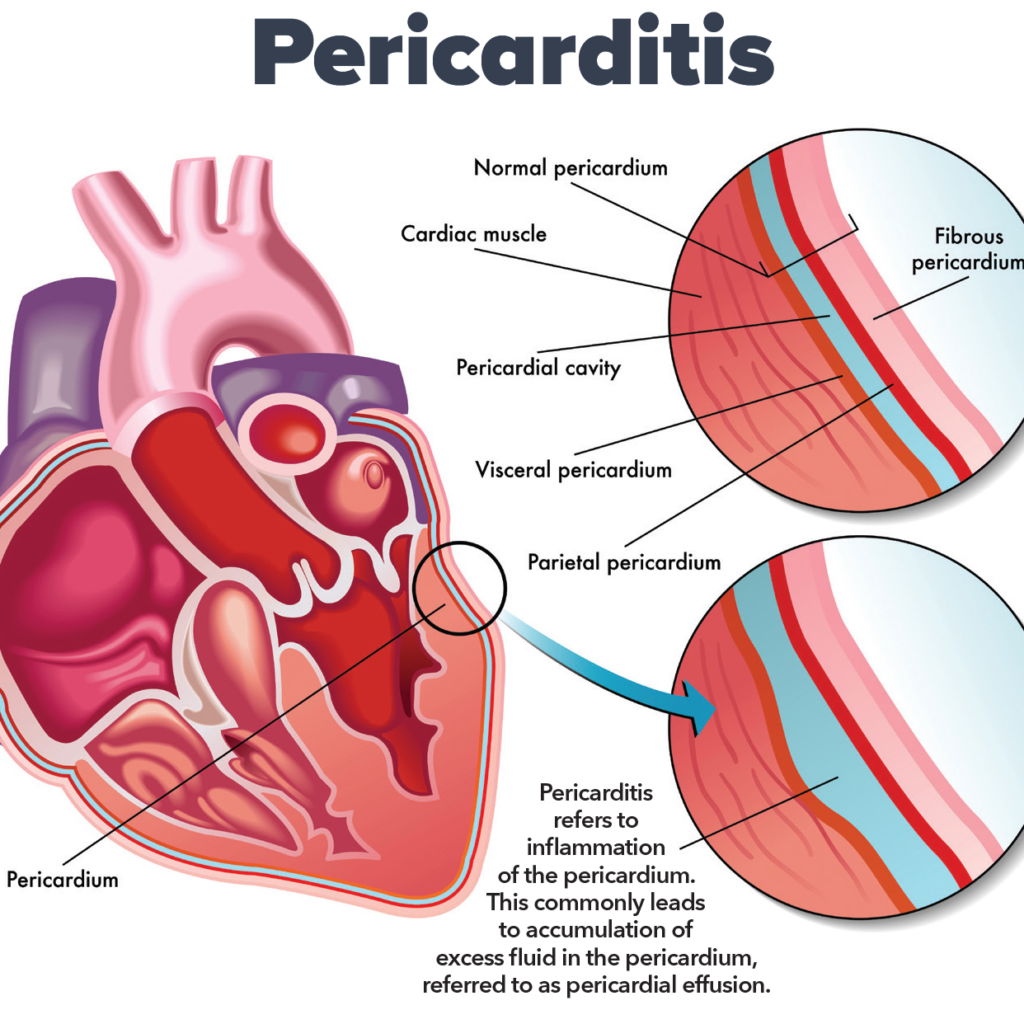
INTRODUTION: What is pericardium ? Term pericardium is derived from the Greek prefix :peri means – Around and Kardia means- Heart pericardium is a double walled fibro serous sac which encloses the heart and roots of great blood vessels. It is situated in middle mediastinum. It consists of 2 layers , they are as follows : STRUCTURE AND FUNCTIONS OF PERICARDIUM: 1.Fibrous Pericardium It is the outer most layer , which encloses the heart and fuses with the roots of great vessels which enter or leave the heart .It is conical sac and made up of dense and loose connective tissue, mainly collagen and elastin .It is non pliable [inflexible] , it can change some what. 1.1 Relations Of Fibrous Pericardium APEX : It is blunt, lies at the level of sternal angle [ angle of louis ] . It fuses with the roots of great vessels and with the pre tracheal fascia. BASE: Broad and inseparably blended with central tendon of the diaphragm. ANTERIOR: It is connected to upper and lower ends of body of sternum [breast bone] by weak superior and inferior sternopericardial ligaments. POSTERIOR It relates to: Each side 1.2 Functions : Protection: As it is made up of dense fibrous tissue it cushions the heart from outside forces and sudden pressure changes . Anchorage: As it is shows attachments with the diaphragm and sternum [ breast bone] it keep the heart in place. Restricting the heart volume : As it is made up of fibrous dense connective tissue .It prevents heart from more expansion than needed. Fibrous prolongations of pericardium: Vessels like aorta, superior vena cava, right and left pulmonary arteries and 4 pulmonary veins receives prolongations of fibrous pericardium up to 5 to 6 mm NOTE: Inferior vena cava doesn’t receive any covering from fibrous pericardium , it enters the pericardium through the diaphragm’s central tendon Protection from infections : Protects heart from infections that might spread from nearby organs like lungs. 2. Serous pericardium : It is a thin double layered serous membrane made up of mesothelium. Parietal layer : This layer fused with the inner surface of fibrous pericardium. Visceral layer: This layer fuses with the heart , except at the cardiac grooves. These two layers, parietal and visceral pericardium continues with each other at roots of great vessels. Pericardial cavity: The space between the parietal and visceral layers is called as pericardial cavity It is filled with pericardial fluid [serous fluid] –15 to 50 ml which is secreted by serous layer. 2.1 Functions: Secretion Of Pericardial Fluid: SINUSES OF PERICARDIUM: Sinuses are the extensions of the pericardial cavity that form between the pericardium and heart surface. There are 3 types of sinuses : TRANSVERSE SINUSES : It is located behind the aorta and pulmonary trunk, and in front of superior vena cava . horizontal gap between arterial and venous ends of heart tube Bounded by : OBLIQUE SINUSES :It is j-shaped narrow space located behind the heart , particularly the left atrium. Bounded by : IMPORTANCE : It permits pulsations of left atrium to take place freely. SUPERIOR SINUSES: It is located in front of ascending aorta and the pulmonary trunk NOTE: it can’t be accessed during electrophysiology procedures. CONTENTS OF PERICARDIUM : BLOOD SUPPLY : Fibrous and parietal layers are supplied by branches of: NERVE SUPPLY: DEVELOPMENT AND CONGENITAL ANOMALIES OF PERICARDIUM: Pericardium embryological development is a complex process that involves several stages, which are intricately linked to the formation of the heart and the cardiovascular system. The pericardium forms from mesodermal layers during embryonic development and undergoes key changes as the heart evolves into its mature anatomical state. 1. Embryological Origins of the Pericardium The pericardium develops from two main mesodermal sources: the somatic mesoderm and the splanchnic mesoderm. The space between these two layers, called the pericardial cavity, is initially a small slit, which later expands and houses the heart. 2. Stages of Pericardium Formation 2.1. Primitive Heart Tube Development (Week 3) At around the third week of embryonic development, the heart begins as a simple, tubular structure formed from the cardiogenic mesoderm. This tube undergoes folding and looping to form a more complex structure, eventually forming the heart with the atria and ventricles. Simultaneously, the pericardial cavity begins to form. Initially, the heart is surrounded by a small amount of mesodermal tissue derived from the lateral mesoderm. The pericardium begins as a continuous sheet of mesoderm surrounding the heart tube. As the heart tube folds, this mesoderm differentiates into the somatic mesoderm (which will form the parietal pericardium) and the splanchnic mesoderm (which will form the visceral pericardium). 2.2. Formation of the Pericardial Cavity (Week 4) By the fourth week, the pericardial cavity becomes more defined. The space between the parietal and visceral layers of mesoderm get enlarged and filled with fluid, allowing the heart tube to move freely within this sac. This fluid-filled pericardial cavity acts as a cushion, reducing friction as the heart begins to beat. 2.3. Development of Pericardial Layers (Week 5 to 6) During weeks 5 and 6, the pericardial cavity enlarges, and the pericardial layers become distinct. The visceral pericardium becomes intimately associated with the developing myocardium (heart muscle), and the parietal pericardium becomes a separate layer, lining the pericardial cavity. At this stage, mesodermal cells in the region of the epicardium (visceral pericardium) also begin to differentiate into epicardial cells, which will contribute to the development of the heart’s coronary vasculature. Meanwhile, the parietal pericardium develops a fibrous structure that will become the fibrous layer of the adult pericardium. 3. Maturation and Differentiation 3.1. Formation of the Fibrous Pericardium The fibrous pericardium is derived from the surrounding mesodermal tissues and becomes an important structure as the heart grows. During the later stages of embryonic development (by week 7-8), the parietal pericardium becomes organized into the fibrous pericardium, which is a tough, inelastic membrane that limits excessive movement of the heart and helps anchor it within the thoracic cavity. 3.2. Pericardial Fluid
Tridosha globeayush.com
Blog

Understanding Concepts 3 Doshas in Ayurveda: Tridosha

1. Introduction Significance of Tridosha in Ayurveda Tridosha, the three fundamental bio-energies—Vata, Pitta, and Kapha—forms the cornerstone of Ayurvedic medicine. It represents the biological energies derived from the five elements (Pancha Mahabhutas: Akasha, Vayu, Agni, Jala, and Prithvi). These energies govern all physiological, psychological, and pathological processes in the human body. Tridosha is the foundation for diagnosing, preventing, and managing diseases in Ayurveda, as it emphasizes restoring balance rather than merely treating symptoms. When in equilibrium, Vata ensures proper movement, Pitta maintains metabolism, and Kapha provides stability. Any disturbance in these doshas leads to disease, showcasing their vital role in maintaining health and wellness. Historical background and foundational principles The concept of Tridosha dates back to the Vedic era, with its earliest mention in texts like the Charaka Samhita and Sushruta Samhita. According to Ayurveda, life is a dynamic interplay of three forces—Vata (wind), Pitta (bile), and Kapha (phlegm)—that work in harmony to sustain life. Each dosha is associated with specific qualities (gunas) and functions. Ancient sages identified how these energies are influenced by diet, lifestyle, seasonal changes, and mental states, creating a unique, personalized approach to health care. The Tridosha theory is based on the idea that the body, mind, and environment are interconnected, and maintaining balance between the doshas leads to optimal well-being. Relevance of Tridosha in modern healthcare In modern healthcare, the Tridosha concept offers a holistic and preventive approach that aligns with personalized medicine. By recognizing individual constitution (Prakriti), Ayurveda provides tailored recommendations for diet, lifestyle, and treatment. Research has highlighted similarities between dosha imbalances and chronic diseases, such as metabolic disorders, stress-related illnesses, and inflammatory conditions. Additionally, the circadian rhythms of doshas correlate with modern chronobiology, emphasizing the role of time in physiological functions. With the growing emphasis on integrative medicine, Tridosha’s principles are gaining recognition for their focus on balance, natural therapies, and promoting long-term health through sustainable lifestyle practices. 2. Nirukti (Etymology) of Tridosha दूषयन्ति इति दोषाः । (A.H.Su. 1:6, Arunadatta) The literary meaning of the word dosha is that which causes decay, destruction or vitiates (others when in aggravated condition). However this is not the correct definition as we all know that when doshas are in balanced condition they carry out all prominent physiological function and maintain health. When doshas get disturbed, they attack other body tissues which are weak and disease is created. The elements, which get spoiled due to vitiated doshas are called as dushya (victims). They are dhatu (body tissue), mala (wastes), organs, channels, sense organs etc. In short dushya are everything in body excluding doshas. Knowledge about dosha is very fundamental in the process of learning Ayurveda. Concept of Tridosha has gradually evolved in Ayurveda, for example – 3. Definition of Tridosha Classical Ayurvedic Definitions The term “Tridosha” is derived from the Sanskrit words “Tri,” meaning three, and “Dosha,” meaning fault or defect. In Ayurvedic texts, “Dosha” is described as a dynamic principle responsible for both the maintenance and disruption of health. According to the Charaka Samhita, Tridosha governs all physiological and psychological functions. Each dosha is made of two primary elements: The equilibrium (samyavastha) of these doshas ensures health, while their imbalance (vikriti) causes disease. Functional Role of Doshas in the Human Body Each dosha plays a distinct role in the functioning of the body: 4. Tridosha: The Three Pillars of Life Tridosha forms the core of Ayurveda and symbolizes the three energies essential for life: Vata, Pitta, and Kapha. These energies control all functions of the body and mind, working together to maintain balance. Vata Dosha: The Energy of Movement Vata represents motion and is linked to air and space. It controls breathing, blood circulation, and all physical and mental movements. When balanced, it supports creativity and flexibility; when imbalanced, it causes restlessness and dryness. Pitta Dosha: The Energy of Transformation Pitta is the energy of heat and digestion, connected to fire and water. It governs metabolism, temperature, and mental focus. Balanced Pitta leads to sharp intellect and a healthy digestive system, but an imbalance can cause anger and inflammation. Kapha Dosha: The Energy of Structure Kapha provides stability and nourishment, linked to earth and water. It maintains body strength, immunity, and emotional calmness. In balance, it ensures growth and stability, while imbalance results in sluggishness and weight gain. 5. Subtypes of Doshas Subtype Specific Function Prana Vata Governs breathing, swallowing, thinking, and sensory perception. Udana Vata Controls speech, memory, energy, and upward movements like exhalation. Samana Vata Regulates digestion, assimilation, and movement of nutrients in the gastrointestinal tract. Vyana Vata Manages circulation, heartbeat, and overall body movements. Apana Vata Oversees elimination processes, such as urination, defecation, and childbirth. Subtypes of Pitta Dosha Subtype Specific Function Pachaka Pitta Responsible for digestion and metabolism in the stomach and intestines. Ranjaka Pitta Influences liver and spleen functions, maintaining blood quality and color. Sadhaka Pitta Supports intelligence, memory, emotional stability, and decision-making. Alochaka Pitta Controls vision and maintains eye health. Bhrajaka Pitta Regulates skin health, complexion, and temperature control. Subtypes of Kapha Dosha Subtype Specific Function Avalambaka Kapha Provides structural stability, supports heart and lung functions. Kledaka Kapha Protects the stomach lining, aids digestion, and maintains lubrication. Bodhaka Kapha Regulates taste perception and saliva production in the mouth. Tarpaka Kapha Nourishes and lubricates the brain and spinal cord for better mental health. Shleshaka Kapha Lubricates joints, supports smooth movement, and strengthens the skeletal system. 6. Tridosha in Diagnosis Diagnosis of Tridosha Dosha Signs of Imbalance Vata Dry skin, constipation, anxiety, irregular sleep, feeling cold. Pitta Irritable, inflammation, acidity, excessive heat, red complexion. Kapha Lethargy, excess weight, congestion, water retention, sluggish digestion. 7. Scientific Correlations with Tridosha Modern Interpretations of Dosha Functions While Ayurveda is an ancient system of medicine, its principles resonate with contemporary scientific understanding, particularly in the realm of body systems and functions. The doshas—Vata, Pitta, and Kapha—can be understood through modern biological systems and physiological processes. For example, Vata corresponds to the nervous system and movement in the body, as it governs all motion-related functions

Subscribe For Newsletter!

Stay informed with our latest updates and exclusive offers by subscribing to our newsletter. Get first access to new products, special events and essential news. Sign up today to stay connected and never miss out on what’s happening.

You have been successfully Subscribed! Ops! Something went wrong, please try again.

All © 2024 Reserved with GlobeAyush

Scroll to Top