Table of Contents
- INTRODUTION:
- STRUCTURE AND FUNCTIONS OF PERICARDIUM:
- 1.Fibrous Pericardium
- 1.1 Relations Of Fibrous Pericardium
- 1.2 Functions :
- 2. Serous pericardium :
- 2.1 Functions:
- SINUSES OF PERICARDIUM:
- CONTENTS OF PERICARDIUM :
- BLOOD SUPPLY :
- NERVE SUPPLY:
- DEVELOPMENT AND CONGENITAL ANOMALIES OF PERICARDIUM:
- Clinical Anatomy :
- Conclusion :
- Reference :
- Read other articles
INTRODUTION:
What is pericardium ?
Term pericardium is derived from the Greek prefix :peri means – Around and Kardia means- Heart
pericardium is a double walled fibro serous sac which encloses the heart and roots of great blood vessels. It is situated in middle mediastinum.
It consists of 2 layers , they are as follows :
- Fibrous pericardium
- Serous pericardium

STRUCTURE AND FUNCTIONS OF PERICARDIUM:
1.Fibrous Pericardium
It is the outer most layer , which encloses the heart and fuses with the roots of great vessels which enter or leave the heart .It is conical sac and made up of dense and loose connective tissue, mainly collagen and elastin .It is non pliable [inflexible] , it can change some what.
1.1 Relations Of Fibrous Pericardium
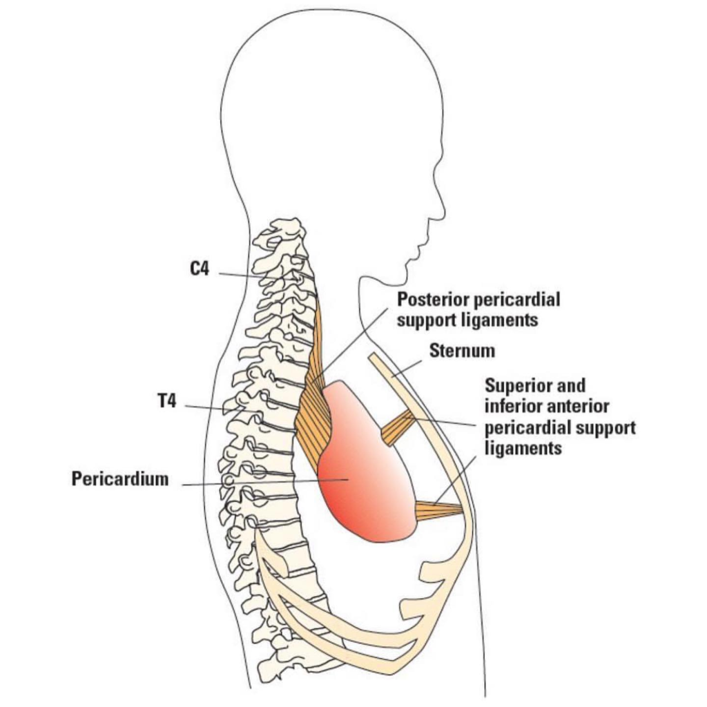
APEX : It is blunt, lies at the level of sternal angle [ angle of louis ] . It fuses with the roots of great vessels and with the pre tracheal fascia.
BASE: Broad and inseparably blended with central tendon of the diaphragm.
ANTERIOR: It is connected to upper and lower ends of body of sternum [breast bone] by weak superior and inferior sternopericardial ligaments.
POSTERIOR
It relates to:
- Principal bronchi
- Oesophagus
- Descending thoracic aorta
- Nerve plexuses around it.
Each side
- Mediastinal pleurae
- Mediastinal surface of lung
- Phrenic nerve
- Pericardiacophrenic vessels


1.2 Functions :
Protection:
As it is made up of dense fibrous tissue it cushions the heart from outside forces and sudden pressure changes .
Anchorage:
As it is shows attachments with the diaphragm and sternum [ breast bone] it keep the heart in place.
Restricting the heart volume :
As it is made up of fibrous dense connective tissue .It prevents heart from more expansion than needed.
Fibrous prolongations of pericardium:
Vessels like aorta, superior vena cava, right and left pulmonary arteries and 4 pulmonary veins receives prolongations of fibrous pericardium up to 5 to 6 mm
NOTE: Inferior vena cava doesn’t receive any covering from fibrous pericardium , it enters the pericardium through the diaphragm’s central tendon
Protection from infections :
Protects heart from infections that might spread from nearby organs like lungs.
2. Serous pericardium :
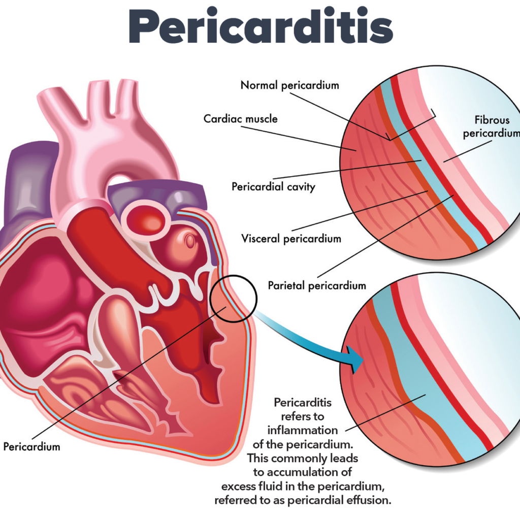
It is a thin double layered serous membrane made up of mesothelium.
- outer : parietal layer
- inner : visceral layer
Parietal layer : This layer fused with the inner surface of fibrous pericardium.
Visceral layer: This layer fuses with the heart , except at the cardiac grooves.
These two layers, parietal and visceral pericardium continues with each other at roots of great vessels.
Pericardial cavity: The space between the parietal and visceral layers is called as pericardial cavity
It is filled with pericardial fluid [serous fluid] –15 to 50 ml which is secreted by serous layer.

2.1 Functions:
Secretion Of Pericardial Fluid:
- Lubrication: fluid secretion helps in reducing friction and protect it from injury.
- Protection : prevents from over distending.
- Mechanical protection: it lubricates and reduces the friction and give mechanical protection to heart and allow heart beat smoothly.
SINUSES OF PERICARDIUM:
Sinuses are the extensions of the pericardial cavity that form between the pericardium and heart surface.
There are 3 types of sinuses :
- Transverse sinuses
- oblique sinuses
- superior sinuses
TRANSVERSE SINUSES : It is located behind the aorta and pulmonary trunk, and in front of superior vena cava .
horizontal gap between arterial and venous ends of heart tube
Bounded by :
- Anteriorly : Ascending aorta and pulmonary trunk
- Posteriorly :Superior vena cava
- Inferiorly : Left atrium
- Each side : Opens into general pericardial cavity .

OBLIQUE SINUSES :It is j-shaped narrow space located behind the heart , particularly the left atrium.
Bounded by :
- Anterior : Left atrium
- Posterior : Parietal pericardium and oesophagus
- On Right and Left :Reflections of pericardium
- Below and Left : Opens into rest of pericardial cavity.
IMPORTANCE : It permits pulsations of left atrium to take place freely.

SUPERIOR SINUSES: It is located in front of ascending aorta and the pulmonary trunk
NOTE: it can’t be accessed during electrophysiology procedures.
CONTENTS OF PERICARDIUM :
- Heart and cardiac vessels and nerves.
- Ascending aorta
- Pulmonary trunk
- Lower half of superior vena cava
- Terminal part of the pulmonary veins .
BLOOD SUPPLY :
Fibrous and parietal layers are supplied by branches of:
- Internal thoracic
- Musculophrenic arteries
- Descending thoracic aorta
- Veins drains into corresponding veins.
NERVE SUPPLY:
- Fibrous and parietal pericardium – phrenic nerves [ pain sensitive ]
- Epicardium : Autonomic nerves of heart [ Not pain sensitive ]

DEVELOPMENT AND CONGENITAL ANOMALIES OF PERICARDIUM:
Pericardium embryological development is a complex process that involves several stages, which are intricately linked to the formation of the heart and the cardiovascular system. The pericardium forms from mesodermal layers during embryonic development and undergoes key changes as the heart evolves into its mature anatomical state.
1. Embryological Origins of the Pericardium
The pericardium develops from two main mesodermal sources: the somatic mesoderm and the splanchnic mesoderm.
- Somatic Mesoderm: This layer contributes to the formation of the parietal pericardium, the outer layer of the pericardial sac.
- Splanchnic Mesoderm: This layer forms the visceral pericardium (or epicardium), which directly covers the heart and great vessels.
The space between these two layers, called the pericardial cavity, is initially a small slit, which later expands and houses the heart.
2. Stages of Pericardium Formation
2.1. Primitive Heart Tube Development (Week 3)
At around the third week of embryonic development, the heart begins as a simple, tubular structure formed from the cardiogenic mesoderm. This tube undergoes folding and looping to form a more complex structure, eventually forming the heart with the atria and ventricles.
Simultaneously, the pericardial cavity begins to form. Initially, the heart is surrounded by a small amount of mesodermal tissue derived from the lateral mesoderm. The pericardium begins as a continuous sheet of mesoderm surrounding the heart tube. As the heart tube folds, this mesoderm differentiates into the somatic mesoderm (which will form the parietal pericardium) and the splanchnic mesoderm (which will form the visceral pericardium).
2.2. Formation of the Pericardial Cavity (Week 4)
By the fourth week, the pericardial cavity becomes more defined. The space between the parietal and visceral layers of mesoderm get enlarged and filled with fluid, allowing the heart tube to move freely within this sac. This fluid-filled pericardial cavity acts as a cushion, reducing friction as the heart begins to beat.
2.3. Development of Pericardial Layers (Week 5 to 6)
During weeks 5 and 6, the pericardial cavity enlarges, and the pericardial layers become distinct. The visceral pericardium becomes intimately associated with the developing myocardium (heart muscle), and the parietal pericardium becomes a separate layer, lining the pericardial cavity.
At this stage, mesodermal cells in the region of the epicardium (visceral pericardium) also begin to differentiate into epicardial cells, which will contribute to the development of the heart’s coronary vasculature. Meanwhile, the parietal pericardium develops a fibrous structure that will become the fibrous layer of the adult pericardium.
3. Maturation and Differentiation
3.1. Formation of the Fibrous Pericardium
The fibrous pericardium is derived from the surrounding mesodermal tissues and becomes an important structure as the heart grows. During the later stages of embryonic development (by week 7-8), the parietal pericardium becomes organized into the fibrous pericardium, which is a tough, inelastic membrane that limits excessive movement of the heart and helps anchor it within the thoracic cavity.
3.2. Pericardial Fluid and Function
As the heart grows and the pericardial cavity continues to expand, the pericardial fluid inside the cavity increases, ensuring that the heart can contract and expand with minimal friction. The epicardial layer (visceral pericardium) also produces this fluid, which helps to lubricate the heart and prevent damage from friction.
3.3. Changes During Fetal Development
By the second trimester, the pericardium reaches its mature structure, with the pericardial cavity, parietal pericardium, and visceral pericardium well-defined. The parietal pericardium is connected to the diaphragm and the sternum through ligaments that provide stability to the heart within the thoracic cavity.
4. Postnatal Changes and Adult Pericardium
After birth, the pericardium maintains its protective and lubricating role. However, during postnatal life, some changes in its structure and function can occur, particularly in response to changes in the heart’s activity, position, and the mechanical forces acting on it.
- Parietal Pericardium: The fibrous layer remains as a tough, dense connective tissue structure that helps limit heart expansion.
- Visceral Pericardium: The visceral pericardium (epicardium) is closely attached to the heart and is involved in the formation of the coronary blood vessels, playing a key role in cardiac vascularization.
In some individuals, the pericardial cavity may also develop adhesions or other anomalies, such as pericardial effusion, which can be related to infections or inflammation.
5. Congenital Anomalies of the Pericardium
In rare cases, abnormalities can occur during the development of the pericardium, including:
- Congenital absence of the pericardium: This is a rare condition where one or more layers of the pericardium are missing. It can lead to the heart being more mobile within the thoracic cavity.
- Pericardial cysts: These are fluid-filled sacs that can form within the pericardium.
- Pericardial adhesions: Abnormal connections can form between the pericardium and surrounding structures.
These anomalies can result in clinical symptoms, including chest pain, shortness of breath, or difficulty with heart function, depending on the nature of the anomaly.

Clinical Anatomy :
- Acute Pericarditis : inflammation of pericardium.
- Pericardial effusion : excessive fluid in the pericardial sac .
- Cardiac tamponade : pressure on the heart that occurs when blood or fluid is filled in between the heart muscles and the outer covering sac [ Pericardium ]
- Chronic constructive pericarditis : pericardium becomes stiffer and thicker than normal.


Conclusion :
The pericardium is a crucial structure in the human cardiovascular system, providing essential protection, lubrication, and support to the heart. Its development begins early in embryogenesis from mesodermal tissues, forming distinct layers -the parietal and visceral pericardium , around the growing heart. The pericardial cavity, which forms between these layers, allows the heart to move freely while minimizing friction. As development progresses, the fibrous pericardium becomes established, stabilizing the heart within the thoracic cavity. This intricate process ensures the heart’s functionality and minimizes the risk of injury or damage during cardiac movements. Despite its structural simplicity, the pericardium plays a vital role in heart protection and overall cardiovascular health. Understanding its embryology helps in diagnosing and managing congenital anomalies and offers insights into heart-related diseases and treatments.
Reference :
- The Pericardium’s Role in Biomechanics
Explains the mechanical function of the pericardium in heart pumping and its interactions with myocardial tissue.
Read more here. - Structure and Function in Health and Disease
Covers pericardial anatomy, its protective role, and its impact on diseases like pericarditis.
Read more here. - Pericardium Anatomy and Physiology
Focuses on anatomical layers, immune functions, and post-surgical healing processes.
Read more here. - Interventions and Therapeutics
Discusses clinical approaches for pericardial diseases and relevant surgical techniques.
Read more here. - Essential Anatomy for Clinical Use
Detailed anatomical analysis for medical procedures like biopsies and pericardioscopy.
Read more here.
Read other articles
- Global Perspectives on Folliculitis: Ayurveda’s Approach to Prevention, Treatment, and Healthcare Disparities
- How to Lose Weight in 1 Week: Ayurvedic and Research-Based Insights
- 50 Research Paper Insights into Sleep: The Ultimate Guide to Health, Cognitive Performance, and Longevity
- Top 20 Most comely Useful Instruments in Physiology lab with their classification- part 4
- BEST AYURVEDIC DIET AND NUTRITION GUIDE
- AYURVEDA INTRODUCTION
- Growth of the Ayurveda Wellness Market in 2024: Personalization, Technology, and Global Expansion
- Shat Kriyakala: Understanding 6 Stage of Disease Progression in Ayurveda and Its Modern Relevance
- Understanding Concepts 3 Doshas in Ayurveda: Tridosha
- Anatomy, Function, and Clinical Insights into the Mediastinum: A Comprehensive Overview


