Table of Contents
INTRODUCTION:
Mediastinum: The mediastinum is the central compartment of the thoracic cavity, located between the lungs.
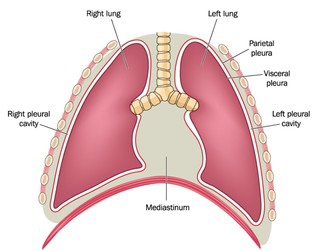
What is the mediastinum?
(MEE-dee-uh-STY-num) The area between the lungs. The organs in this area include the heart and its large blood vessels, the trachea, the esophagus, the thymus, and lymph nodes but not the lungs.
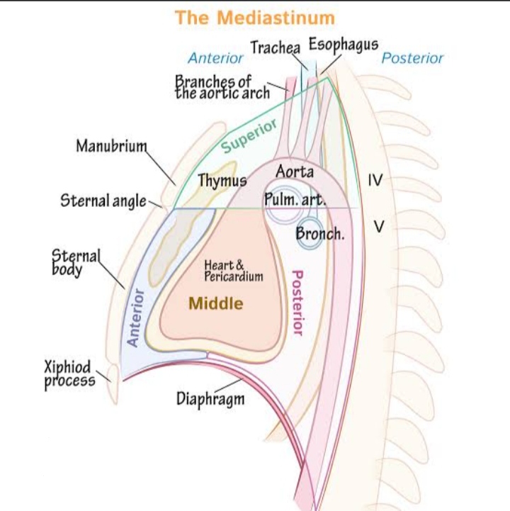
It is divided into : Superior and Inferior compartments.
GENERAL BOUNDARIES OF MEDIASTINUM:
- ANTERIOR: Sternum
- POSTERIOR: Vertebral column
- SUPERIOR: Thoracic inlet
- INFERIOR : Diaphragm
- LATERAL SIDES : Mediastinum Pleura
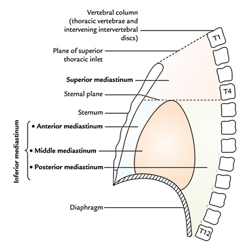
SUPERIOR MEDIASTINUM:
BOUNDARIES :
- ANTERIOR: Manibrium sterni .
- POSTERIOR: Thoracic vertebrae (T1 to T4).
- SUPERIOR: Thoracic inlet
- INFERIOR : Imaginary plane passing from sternal angle (Angle of luis) to lower border of T4 Vertebrae.
- ON EACH SIDE (Lateral): Medistinal pleura.
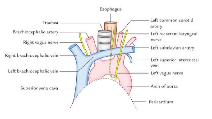
CONTENTS :
1.Trachea
2. Oesophagus
3.Arch of aorta and its 3 branches:
- left subclavian artery
- Brachiocephalic artery
- Left common carotid artery.
4. Superior venacava(upper half) and its branches
- Left brachiocephalic vein
- Right brachiocephalic vein
5. MUSCLES :
- Sternohyoid
- Sternothyroid
- Lower end of longus colli.
NERVES :
- Vagus nerve
- Phrenic nerves
- Cardiac nerves from both sides
- Left recurrent nerve
- Laryngeal nerve.
Lymphatics :
- Thymus
- Thoracic duct
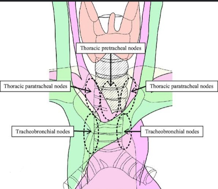
- Lymph nodes- para tracheal
- brachiocephalic
- tracheobronchial.
INFERIOR MEDIASTINUM
it is divided into 3 parts :
1.Anterior
2.Middle
3.Posterior
ANTERIOR MEDIASTINUM:
it is a narrow space present infront of pericardium of heart and behind the body of sternum. It is overlapped by thin Anterior border of both lungs.
BOUNDARIES:
- Anterior – body of sternum
- Posterior – pericardium
- Superior – imaginary line separating superior and inferior Mediastinum.
- Inferior – anterior part of superior surface of diaphragm.
- On each side – mediastinal Pleura.
CONTENTS:
1.Sternopericardial ligament
2. Areolar tissue
3. Lowest part of Thymus.
4. Small mediastinal branches of internal thoracic artery.
5. Lymph nodes.
MIDDLE MEDIASTINUM:
it is the place where heart inside pericardium and its contents are present.
Boundaries:
- Anterior: anterior Mediastinum
- Posterior: Posterior Mediastinum , Azygous vein.
- Superior: imaginary line separating superior and inferior Mediastinum.
- Inferior: diaphragm
- Sides : Mediastinal Pleura
CONTENTS
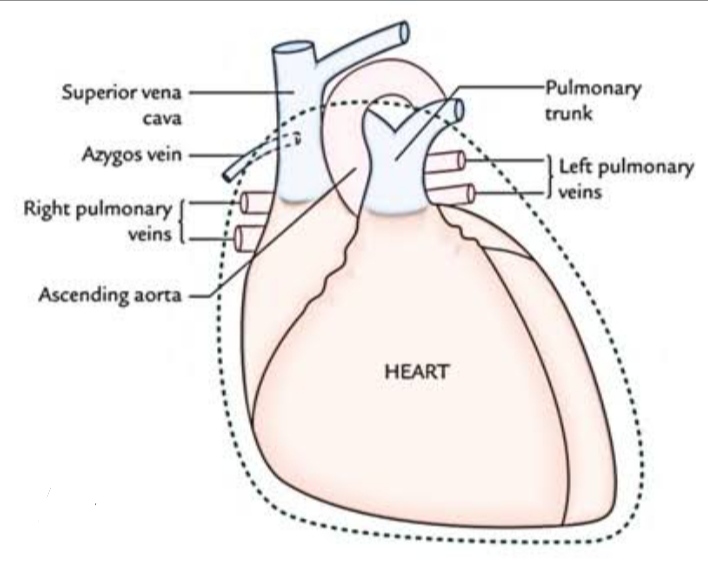
- Heart
- azygous vein
- ascending aorta
- left and right pulmonary veins
- lower part of superior venacava
- pulmonary trunk
- Nerves – phrenic,deep cardiac plexus
- lymph nodes – Tracheobronchial nodes
- pericardium enclosing heart.
POSTERIOR MEDIASTINUM :
it is present behind the pericardium of heart and infront of vertebral column.
BOUNDARIES :
- Anterior: Trachea bifurcation,pulmonary vessels,pericardium
- posterior: vertebrae(T4- T8) and intervening discs.
- superior : imaginary line separating superior and inferior Mediastinum.
- Inferior: Posterior part of upper surface of diaphragm.
CONTENTS
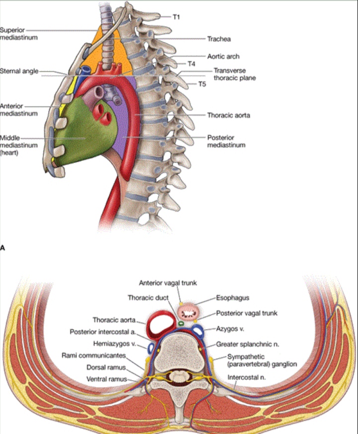
- Oesophagus
- descending thoracic aorta and its branches.
- azygous vein
- Accessory hemiazygous vein
- Hemiazygous vein
- Nerves: vagus , splanchic nerves(greater,lesser,least from lower 8th thoracic ganglia of sympathetic chain)
- Thoracic duct.
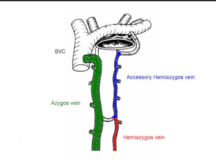
FUNCTIONS OF MEDIASTINUM:
- Protection : It protects heart, thymus, trachea,oesophagus,lymph nodes etc.
- passage way: It acts as passage way for digestion and respiration as oesophagus and trachea pass through it.
- Blood circulation : It keeps heart in place and help heart to pump blood .
- It acts as house of heart and roots of great vessels.
CLINICAL ANATOMY:
- The prevertebral layer of the deep cervical fascia extends to the superior mediastinum, and is attached to the fourth thoracic vertebra(T4). An infection present in the neck behind this fascia can pass down into the superior mediastinum but not lower down.
- The pretracheal fascia of the neck also extends to the superior mediastinum, where it blends with the arch of the aorta. Neck infections between the pretracheal and prevertebral fasciae can spread into the superior mediastinum, and through it into the posterior mediastinum. Thus mediastinitis can result from infections in the neck .
- There is very little loose connective tissue between the mobile organs of the mediastinum. Therefore,the space can be readily dilated by inflammatory fluids, neoplasms, etc.
- In the superior mediastinum, all large veins are on the right side and the arteries on the left side. During increased blood flow veins expand enormously, while the large arteries do not expand at all . Thus there is much ‘dead space’ on the right side and it is into this space that tumour or fluids of the mediastinum tend to project
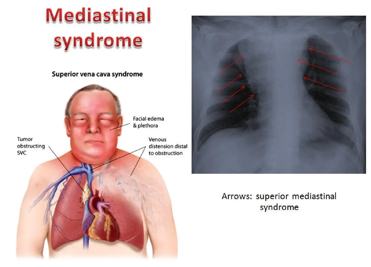
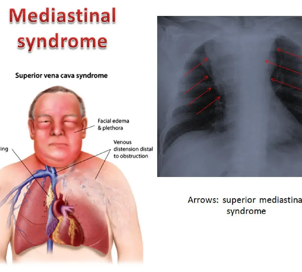
- Compression of mediastinal structures by any tumour gives rise to a group of symptoms known as mediastinal syndrome. The common symptoms are as follows.
- a.Obstruction of superior vena cava gives rise to engorgement ( swelling ) of veins in the upper half of the body.
- b. Pressure over the trachea causes dyspnoea(shortness of breath ) and cough.
- c. Pressure on oesophagus causes dysphagia ( difficulty in swallowing ).
- d. Pressure on the left recurrent laryngeal nerve gives rise to Hoarseness of voice (dysphonia).
- e. Pressure on the phrenic nerve causes paralysis of the diaphragm on that side.
- f. Pressure on the intercostal nerves gives rise to pain in the area supplied by them. It is called intercostal neuralgia.
- g. Pressure on the vertebral column may cause erosion of the vertebral bodies.
- The common causes of mediastinal syndrome are bronchogenic carcinoma, Hodgkin’s lymphoma (HL) disease causing enlargement of the mediastinal lymph nodes,aneurysm or dilatation of the aorta, etc.
References
- NCBI – Detailed anatomy, compartments, and clinical relevance of the mediastinum. Read more.
- SEER Training – Overview of mediastinal structures like the heart, trachea, and esophagus. Visit here.
- University of Minnesota – Development and anatomy of the mediastinum from an embryological perspective. Learn more.
- Merck Manual – Summary of mediastinal compartments and clinical conditions. Details here.
- TeachMeAnatomy – Anatomy and functions of the mediastinum with diagrams. Explore here.
Read other articles
- How to Lose Weight in 1 Week: Ayurvedic and Research-Based Insights
- 50 Research Paper Insights into Sleep: The Ultimate Guide to Health, Cognitive Performance, and Longevity
- Top 20 Most comely Useful Instruments in Physiology lab with their classification- part 4
- BEST AYURVEDIC DIET AND NUTRITION GUIDE
- AYURVEDA INTRODUCTION
- Growth of the Ayurveda Wellness Market in 2024: Personalization, Technology, and Global Expansion
- Shat Kriyakala: Understanding 6 Stage of Disease Progression in Ayurveda and Its Modern Relevance
- GET DISEASE FREE LIFE: 8 EXCEPTIONAL FEATURES OF HEALTHY PEOPLE THAT SET THEM DISEASE FREE: Ayurvedic Edition

