
Introduction :
Kidneys are a pair of retroperitoneal [covered only on anterior side by peritoneum] organs which are part of excretory system.
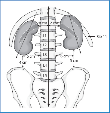
Location : Situated on the posterior abdominal wall behind the peritoneum, one on each side of vertebral column . It covers epigastric , hypogastric ,lumbar and umbilical regions of abdomen .
In vertical view : It covers upper border of 12th Thoracic vertebrae [T12] to center body of 3rd Lumbar vertebrae.
NOTE : Right kidney is lower than the Left kidney due to presence of liver on right side . The transpyloric line pass through the upper part of hilus of right kidney and lower part of hilus of left kidney respectively.
General Features of Kidney:
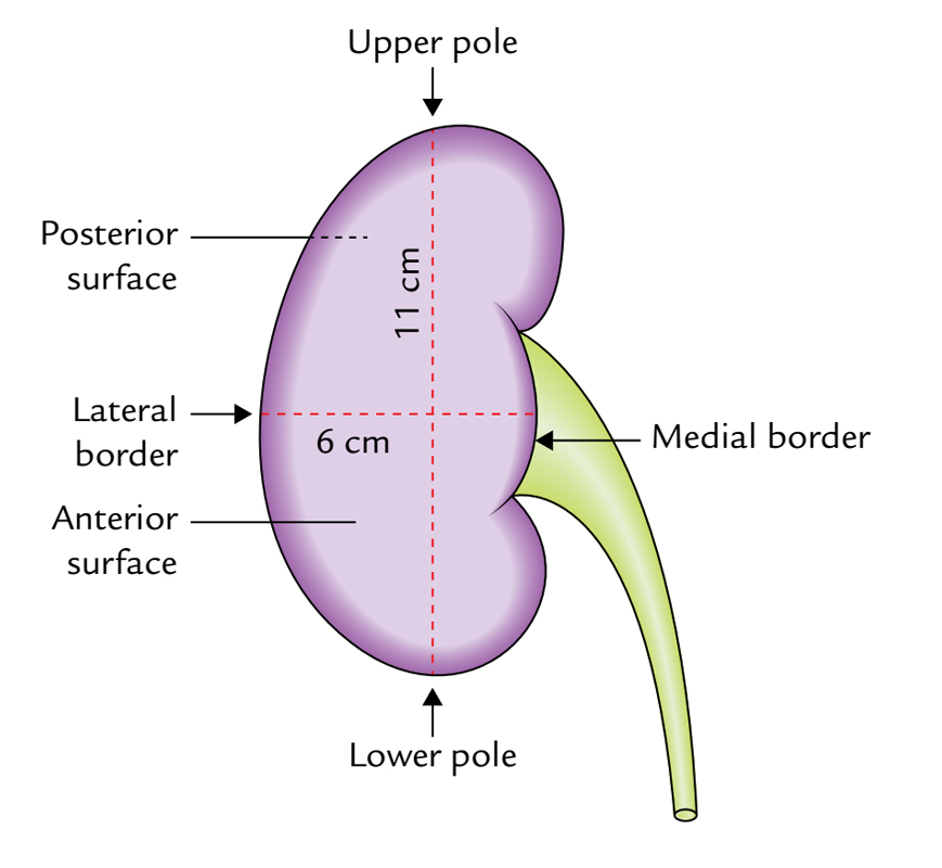
- Shape : Bean shape
- Size : 11cm length
- 6cm broad
- 3cm thick
- Weight : Males – 150gms
- Females-135gms
- Color : Reddish brown
:
External Features of Kidney :
It consists of :
2 poles :
- Upper pole– close contact with adrenal gland[suprarenal gland]
- Lower pole -pointed
2 surfaces :
- Anterior : Irregular
- Posterior : Flat
NOTE : It is difficult to recognize anterior and posterior surfaces of kidney .
2 borders :
- Lateral border : Convex
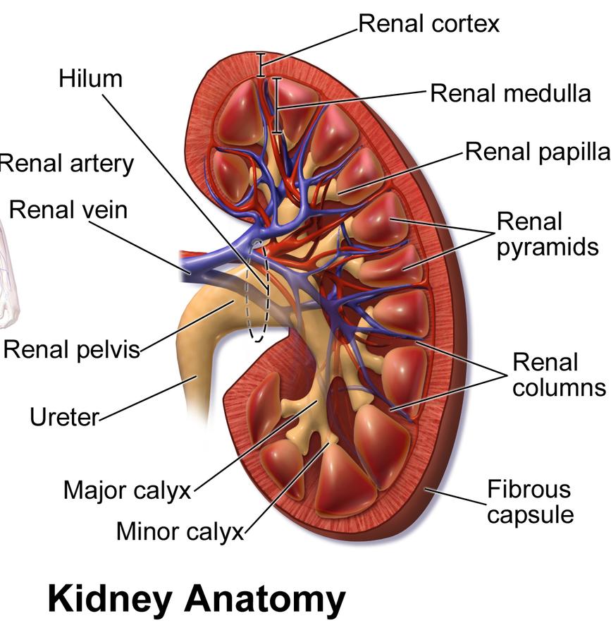
- Medial border : Concave [ it shows depression called as hilum ]
structures coming in and going out of the hilum from anterior to posterior side .
- Renal vein
- Renal artery
- Renal pelvis [ This is expanded part of ureter ]
Some times , Renal artery may enter hilus behind Renal pelvis and Renal vein tributary may also found in same plane that of renal pelvis.
Relations Of Kidney :
Relations common in both kidney .
- Upper pole : Adrenal gland [Supra renal gland]
- Lower pole : It is 2.5 cm above iliac crest
- Medial border : hilus
- above hilus – adrenal gland
- below hilus – ureter
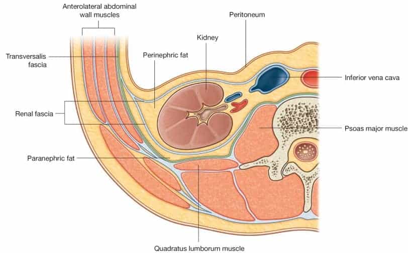
- Anterior : peritoneum [retroperitoneal organ]
- Posterior : diaphragm
- Medial and lateral accurate ligaments
- Psoas major muscle
- Quadratus lumborum muscle
- Transverse abdominis
- Vessels – sub costal vessels
- Nerves – sub costal iliohypogastric and ilioinguinal nerves.
- Hilum : Renal artery
- Renal vein
- Renal pelvis.

Other relations of kidneys :
Right kidney :
- Right supra renal gland
- liver [right lobe]
- Hepatic flexure of colon
- Small intestine
- 2nd part of duodenum
NOTE :
- The right lobe and hepatic flexure of colon are related to lateral kidney
- Hepatic flexure of colon and small intestine are covered by peritoneum.
Left kidney:
- Left suprarenal gland
- spleen
- splenic vessels
- splenic flexure
- descending colon
- pancreas
- stomach
- jejunum
NOTE:
- Gastric , splenic, and jejunal parts are covered by peritoneum
- Spleen and Descending colon related to lateral border of Left kidney.

Coverings of kidney [Capsules] :
There are 4 coverings of kidney :
- Fibrous capsule
- perirenal or perinephric fat
- Renal fascia
- Pararenal or paranephric fat
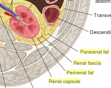
Fibrous capsule : It is thin membrane ,closely associate with kidney and lines renal sinus . It is smooth and convex . It can be easily stripped off. But in certain diseases it becomes adherent and cannot stripped off.
Perirenal or Perinephric fat : It is a adipose tissue which lines the fibrous capsule ,and it is thickest at borders of kidney and fills up extra space in the renal sinus. It extends over adrenal gland superiorly.
Nephroptosis [Floating kidney] : It is the condition in which the absence of perirenal fat or seen in severe loss of weight.
Renal Fascia [Gerota’s fascia] :
It surrounds the perirenal fat and suprarenal glands [Adrenal gland] .
This renal fascia is divided into 2 layers :
- Anteriorly : It is called as Fascia of Toldt . In some texts it is consider as Gerota’s fascia
- Posteriorly : It is called as Fascia of Zuckerkandl
A number of trabeculae connect the renal fascia to fibrous capsule across perirenal fat.
Position of renal fascia :
TRACED ABOVE :
Two layers meet at the upper pole of kidney and split to enclose suprarenal gland and meet again at upper pole of suprarenal gland continue with fascia covering diaphragm.
TRACED BELOW :
These two layers remain separate and encloses ureter :
- Anteriorly Layer : It fuses with extra peritoneal fatty tissue at right iliac fossa
- Posterior Layer : This layer blended with fascia iliaca.
LATERALLY:
Laterally these layers fuses together and continued with the fascia taransversalis behind colon.
MEDIALLY :
- Anterior layer : This layer passes in front of renal vessels and it continues with fascia covering abdominal aorta and inferior vena cava .
- Posterior layer : This layer covers quadratus lumborum and psoas major muscle.
- Medially these two layers of fascia connect through septum , and the renal vessels pierces this septum and enters the hilum of kidney.
Pararenal fascia :
It is present dorsal to renal fascia , this is more on posterior side to fill paravertebral space and acts cushion.
Internal structure of kidney :
- Consists of following :
- Outer [cortex] : It is reddish brown in color
- Inner [medulla] : pale in color
- Renal sinus
CORTEX : It consists of two parts :
- Cortical arches or cortical lobules : this part of cortex form caps over the bases of renal pyramid
- Renal columns : This is also called as column’s of bertini’s . This is the part of cortex extends in between the pyramids of medulla .
MEDULLA :
It is made up of conical masses called renal pyramids 10 in number in each kidney . These form renal papillae at apices and these indent minor calyces which give rise to major calyces and then into renal pelvis .
RENAL SINUSES :
This is the space extends into kidney from the hilus . It contain
- Branches of renal artery
- Tributaries of renal vein
- Renal pelvis
RENAL PELVIS : This part gives rise to 2 to 3 major calyces and these give rise to 7 to 13 minor calyces. these minor calyces ends in an expansion which intended by 1 to 3 renal papillae .
Structure Of Uriniferous Tubule
There are 1 to 3 million tubules in each kidney . These are the structural and functional units of kidney.
Each tubule consists of two parts :
- Excretory part
- Collecting part
Excretory part : This part of tubule is called as nephron
- This part includes :
- Renal corpuscle or Malpighian corpuscle : This part consists of Bowman’s capsule and a tuft of capillaries entering into the bowman’s capsule called as glomerulus .
- FUNCTION : Filtration of substances from plasma
- Renal Tubule :It consists of
- Proximal convoluted tubule [PCT]
- Loop of Henle’s [LOH]
- Distal convoluted tubule [DCT]
- FUNCTION : Selective resorption
- Renal corpuscle or Malpighian corpuscle : This part consists of Bowman’s capsule and a tuft of capillaries entering into the bowman’s capsule called as glomerulus .
Collecting part :
This is the part where all filtrate finally reaches , this is called as Collecting duct . Many collecting ducts unite and form Ducts of bellini which opens into minor calyces through renal papillae.

Juxta Glomerular Apparatus
The Juxtaglomerular Apparatus (JGA) are specialized structures in the kidneys that plays a critical role in regulating blood pressure, filtration rate, and electrolyte balance.
It is located near the junction of the afferent arteriole and the distal convoluted tubule (DCT) of a nephron. The JGA helps maintain homeostasis, particularly by controlling the renin-angiotensin-aldosterone system (RAAS), which regulates blood volume, blood pressure, and sodium balance.
Components of the Juxtaglomerular Apparatus:
- Macula Densa:
- The macula densa is a group of specialized cells in the distal convoluted tubule, located close to the glomerulus. These cells are sensitive to the sodium chloride (NaCl) concentration in the filtrate.
- When the sodium concentration in the tubular fluid is low, the macula densa signals the JGA to increase the release of renin, which helps restore normal sodium balance and blood pressure.
- Juxtaglomerular Cells (Granular Cells):
- These are modified smooth muscle cells in the wall of the afferent arteriole (and sometimes the efferent arteriole). They are called granular cells due to their cytoplasmic granules containing renin.
- When the macula densa detects low sodium levels or low blood pressure (through baroreceptors in the afferent arteriole), the granular cells release renin into the bloodstream. Renin is the first enzyme in the RAAS pathway.
- Extraglomerular Mesangial Cells (Lacis Cells):
- These are specialized cells found between the macula densa and the afferent/efferent arterioles. They are involved in structural support and communication between the macula densa and the granular cells.
- The exact role of these cells is still not completely understood, but they are thought to assist in signal transduction during the regulation of blood flow and renin release.

Blood supply :
Vascular Segments :
- Renal artery is divided into 5 segments :
- 4 anterior segments
- 1 posterior segments
BLOOOD SUPPLY :

Lymphatic Drainage :
- Lateral aortic nodes
- Location : At origin of renal arteries
Clinical anatomy
- Nephritis : Inflammation of kidney
- pyelonephritis : Inflammation of kidney due to bacterial infection.
- Tuberculosis of kidney : It is caused by the same mycobacterium tuberculosis which causes lung tuberculosis. It shows symptoms like blood in urine , Flank pain ,Discomfort etc.
- Tumors : Abnormal growth of kidney
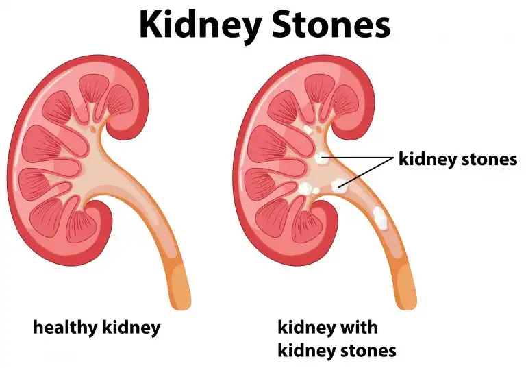
- Urine get concentrated in kidney leads to kidney stones [stag horn stones].
- Increased urea in blood indicates suppressed kidney function.
- Chronic renal failure : In this case a procedure called dialysis is done .
- The kidneys are likely to be injured due to penetrating injuries to lower thoracic cage. These may also be injured by kicks in the renal angle between the vertebral column and 12th rib.
- The angle between the lower border of the 12th rib and the outer border of the erector spinae is known as the renal angle. It overlies the lower part of the kidney. Tenderness in the kidney is elicited by applying pressure over this angle, with the thumb .
- Blood from a ruptured kidney or pus in a perinephric abscess first distends the renal fascia, then forces its way within the renal fascia downwards into the pelvis. It cannot cross to opposite side because of the fascial septum and midline attachment of the renal fascia.
- In surgical exposures of the kidney, when sometimes the 12th rib is resected for easier delivery of the kidney, danger of opening the pleural cavity must be borne in mind. The lower border of the pleura lies in between the 12th rib and the diaphragm. The order of structures from anterior to posterior side being diaphragm, pleura and rib.
- When the 12th rib is absent or is too short to be felt, the 11th rib may be mistaken for the 12th, and the chances of opening the pleural cavity are greatly increased . Lithotripsy has replaced conventional method to some degree.
NOTE : Kidney stones lie on the body of vertebrae and Gall stones lie on anterior to body of vertebrae.

Conclusion:
In conclusion, the kidneys are vital organs with critical functions in maintaining the body’s internal balance, including waste filtration, fluid and electrolyte regulation, Control blood pressure , and hormone production. Their clinical anatomy, which involves their structure, location, and functional components, is essential for understanding the mechanisms underlying kidney function and the pathophysiology of kidney diseases.
The kidneys’ anatomical features, such as the renal cortex, medulla, nephrons, and blood supply, are closely tied to their physiological roles. Knowledge of kidney anatomy is crucial for diagnosing conditions like chronic kidney disease, acute kidney injury, and kidney stones, as well as for performing medical procedures like biopsies, dialysis, and transplantation. Additionally, understanding congenital and developmental kidney anomalies aids in early diagnosis and management.
Overall, understanding of the kidneys’ anatomy and clinical relevance is fundamental in preventing, diagnosing, and managing renal diseases, ensuring better patient outcomes and quality of life.
References for more details
Websites
- National Kidney Foundation (NKF)
- MedlinePlus – Kidneys
- Healthline – Kidney Anatomy and Function
- PubMed – Kidney Research
- Radiopaedia – Kidney Anatomy
Books
- Human Anatomy and Physiology by Elaine N. Marieb and Katja Hoehn
- Medical Physiology by Guyton and Hall
Our Other articles
- 10 Powerful Meditation Postures to Transform Your Mind and Boost Inner Peace
- Global Perspectives on Folliculitis: Ayurveda’s Approach to Prevention, Treatment, and Healthcare Disparities
- How to Lose Weight in 1 Week: Ayurvedic and Research-Based Insights
- 50 Research Paper Insights into Sleep: The Ultimate Guide to Health, Cognitive Performance, and Longevity
- Top 20 Most comely Useful Instruments in Physiology lab with their classification- part 4
- BEST AYURVEDIC DIET AND NUTRITION GUIDE
- AYURVEDA INTRODUCTION
- Growth of the Ayurveda Wellness Market in 2024: Personalization, Technology, and Global Expansion
- Shat Kriyakala: Understanding 6 Stage of Disease Progression in Ayurveda and Its Modern Relevance
- Understanding Concepts 3 Doshas in Ayurveda: Tridosha
- Anatomy, Function, and Clinical Insights into the Mediastinum: A Comprehensive Overview
- Stress Relief and Mental Wellness: Understanding Causes, Effects and Effective Strategies
- Arthritis explore Ayurvedic and Modern Aspects
- Over view of the Urinary System: Embryology , Functions and Congenital Diseases


Pingback: India’s Ancient Science, Global Validation: Time to Embrace Our Heritage -